
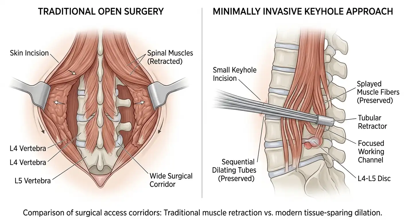
The evolution of the surgical corridor
For many years, the standard way to fix a severely herniated spinal disc was through a large incision in the middle of the back.
To reach the spine, a surgeon had to separate the heavy, supportive back muscles from their bony attachments. This extensive muscle separation was the primary reason patients experienced significant soreness and required long hospital stays after their operations.
Today, advanced instrumentation allows specialists to work through a radically different pathway. Instead of pulling down the entire structural wall to fix a leaky pipe, we work through a tiny window.
This modern shift represents the core philosophy behind minimally invasive lumbar spine surgery mumbai. It focuses on protecting your natural anatomy while clearing the exact point of nerve compression.
How does a slender tube replace a large incision?
If you choose a keyhole discectomy, the procedure begins with a tiny skin incision, often no wider than a standard fountain pen.
Rather than cutting through your lower back muscles, the surgical team uses a series of slender, gently tapered tubes called dilators. These tubes are inserted one over the other, creating a narrow corridor by simply pushing the flexible muscle fibers apart along their natural alignment.
Once the final working tube is securely in place, an ultra-high-definition surgical microscope or an advanced endoscopic camera is attached to the top. This setup floods the tiny spinal workspace with bright, clear light and magnifies the delicate nerve roots up to twenty times their actual size.
Through this narrow tube, micro-instruments are used to gently move the nerve root out of harm’s way and remove only the specific fragment of leaked disc material that was causing your leg agony. The rest of your healthy disc remains untouched and perfectly intact.
Why does muscle preservation change everything?
When the repair is complete, the working tube is carefully withdrawn. Because the muscle fibers were merely separated rather than cut or torn, they snap back together naturally, like the vertical strings of a tennis racket.
This preservation of your structural framework leads to several distinct benefits during your recovery phase.
First, post-operative discomfort is significantly reduced because your back muscles do not have to heal from major traumatic cuts. Second, there is minimal blood loss, which lowers the overall physiological strain on your body.
Finally, because the structural integrity of your lower back remains secure, you are able to stand up, walk, and return home much faster than was ever possible with older surgical methods. Understanding the details of your microdiscectomy recovery timeline can help you plan a smooth, confident return to your normal daily routine.
Is a keyhole approach right for you?
While the benefits of specialized keyhole access are clear, it is important to remember that technology is a tool, not a cure-all. The right approach depends entirely on your specific structural blockage.
If your MRI reveals a simple, localized disc protrusion pressing on a single nerve root, a precise keyhole discectomy is often an excellent choice to achieve rapid, long-lasting relief.
However, if your back pain is caused by widespread bony overgrowth or complex structural instability where multiple bones have slipped out of alignment, a broader structural correction may be required to protect your long-term mobility.
Consult a dedicated spine specialist who can analyze your scans and design a precise treatment plan tailored to your lifestyle. You do not have to let the fear of a large operation keep you living in pain.